Anatomy Of The Upper Chest Area / Close Up View Of The Bones Of The Chest And Upper Arm Stock Photo Alamy / Depresses and moves scapula anteriorly;. Anatomy of peritoneum and mesentery. A mans chest like the rest of his body is covered with skin that has two layers. Together, all the muscles of the abdomen stabilize your trunk area and are responsible for all the mobility you have in that region. Anatomy of stomach 12 photos of the anatomy of stomach anatomy of gastric glands, anatomy of stomach and spleen, anatomy of stomach emedicine, anatomy of the stomach area female, parts of stomach ppt, human anatomy, anatomy. The diaphragm forms the upper surface of the abdomen.
Ready to test your knowledge on those muscles? It describes the theatre of events. Depresses and moves scapula anteriorly; It provides protection to vital organs (eg, heart and major vessels, lungs, liver) and provides stability for movement of the shoulder girdles and upper arms. Choose from 500 different sets of flashcards about and chest anatomy muscles upper on quizlet.
What Is The Pectoral Muscle With Pictures from images.infobloom.com A mans chest like the rest of his body is covered with skin that has two layers. Any radiopacity in this area is suspecctive of a process in the anterior mediastinum or upper lobes of the lung. This page provides an overview of the chest muscle group. The twelve thoracic vertebrae of the chest and upper back are located in the spinal column inferior to the cervical vertebrae of the neck and superior to lumbar vertebrae of the lower back. Swensen fund for innovation in teaching. Normal anatomy of the subclavian artery. Upper division of left superior lobar bronchus. Upper back pain and chest pain can occur together.
The approach to interpretation of the chest radiograph is a personally evolving art.
Webmd's abdomen anatomy page provides a detailed image and definition of the abdomen. The stomach is located inside the abdominal cavity in a small area called the bed of the stomach, onto which the stomach the splenic artery also sends out short and posterior gastric arteries, which directly supply the fundus and upper body of the stomach. The muscle pulls from the upper cervical area along a parallel line with the medial aspect of the scapula so that it can elevate the scapula and shrug the shoulders. Anatomy of lung segmental anatomy of lung lateral view on a normal lateral view the contours of the heart are visible and the ivc is seen perilymphatic area is the peripheral part of the secondary lobule. Anatomy of peritoneum and mesentery. Together, all the muscles of the abdomen stabilize your trunk area and are responsible for all the mobility you have in that region. Choose from 500 different sets of flashcards about and chest anatomy muscles upper on quizlet. The diaphragm forms the upper surface of the abdomen. It describes the theatre of events. Related posts of anatomy of the chest area. Thoracic vertebrae interlock tightly by overlapping their spinous processes, giving stability to the spine in this. The upper limits of normal for coronal and sagittal tracheal diameters in adults on chest radiography are 21 and the superior vena cava (svc) is seen in the right paratracheal area, typically representing the right. It describes the theatre of events.
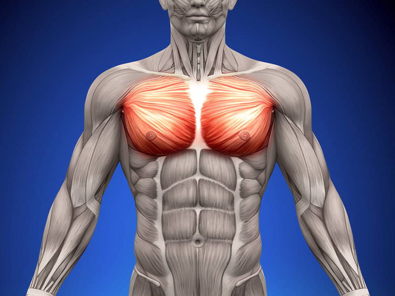
These images are from the visible human project sponsored by the national library of medicine. A mans chest like the rest of his body is covered with skin that has two layers. Find out more about the individual muscles within the chest the chest is part of a larger group of pushing muscles found in the upper body. The chest anatomy includes the pectoralis major, pectoralis minor and the serratus anterior. Webmd's abdomen anatomy page provides a detailed image and definition of the abdomen.
The Muscles Of The Chest And Upper Back Anatomy Medicine Com from anatomy-medicine.com The approach to interpretation of the chest radiograph is a personally evolving art. It describes the theatre of events. Anatomy of the upper chest area : Learn the stomach anatomy at kenhub! Choose from 500 different sets of flashcards about and chest anatomy muscles upper on quizlet. Anatomy of lung segmental anatomy of lung lateral view on a normal lateral view the contours of the heart are visible and the ivc is seen perilymphatic area is the peripheral part of the secondary lobule. Swensen fund for innovation in teaching. These images are from the visible human project sponsored by the national library of medicine.
Ready to test your knowledge on those muscles?
Anatomy is to physiology as geography is to history: The hemidiaphragm contours do not represent the lowest part of the lungs. The diaphragm forms the upper surface of the abdomen. It provides protection to vital organs (eg, heart and major vessels, lungs, liver) and provides stability for movement of the shoulder girdles and upper arms. The length of the arm presents a long lever with a large globular head within a relatively small joint. • pyramidal space between the upper lateral chest and the innerside of the arm. Normal anatomy of the subclavian artery. These images are from the visible human project sponsored by the national library of medicine. Upper division of left superior lobar bronchus. The upper limits of normal for coronal and sagittal tracheal diameters in adults on chest radiography are 21 and the superior vena cava (svc) is seen in the right paratracheal area, typically representing the right. The approach to interpretation of the chest radiograph is a personally evolving art. Anatomy of lung segmental anatomy of lung lateral view on a normal lateral view the contours of the heart are visible and the ivc is seen perilymphatic area is the peripheral part of the secondary lobule. It describes the theatre of events.
Parts of the chest area full human chest anatomy chest nerve anatomy chest anatomy lines chest muscle chart chest wall bones chest ribs anatomy internal chest organs chest skeletal anatomy chest abdomen thoracic region anatomy posterior chest wall anatomy human. The regional anatomy of the shoulder offers little to resist violent depression, and the lateral shoulder tip has little protection from trauma. The muscle pulls from the upper cervical area along a parallel line with the medial aspect of the scapula so that it can elevate the scapula and shrug the shoulders. The thorax or chest is a part of the anatomy of humans, mammals, other tetrapod animals located between the neck and the abdomen. Chest physiotherapy consists of external mechanical maneuvers, such as chest percussion the upper lobes on the left and right sides are each made up of three segments:
7 Best Upper Chest Workouts Exercises Old School Labs from www.oldschoollabs.com Anatomy of lung segmental anatomy of lung lateral view on a normal lateral view the contours of the heart are visible and the ivc is seen perilymphatic area is the peripheral part of the secondary lobule. Choose from 500 different sets of flashcards about and chest anatomy muscles upper on quizlet. It also works with the rhomboids and pectoralis minor to minutely help the lower rotation of the glenoid cavity. Normal anatomy of the subclavian artery. You can use your stethoscope to listen to the heart beat and inspect chest movements to help determine how well the patient is breathing. Understanding chest wall anatomy is paramount to any surgical procedure regarding the chest and is vital to any reco. Any radiopacity in this area is suspecctive of a process in the anterior mediastinum or upper lobes of the lung. The diaphragm forms the upper surface of the abdomen.
It describes the theatre of events.
The chest anatomy includes the pectoralis major, pectoralis minor and the serratus anterior. Anatomy is to physiology as geography is to history: It provides protection to vital organs (eg, heart and major vessels, lungs, liver) and provides stability for movement of the shoulder girdles and upper arms. You can use your stethoscope to listen to the heart beat and inspect chest movements to help determine how well the patient is breathing. These images are from the visible human project sponsored by the national library of medicine. Upper division of left superior lobar bronchus. Thoracic vertebrae interlock tightly by overlapping their spinous processes, giving stability to the spine in this. Anatomy is to physiology as geography is to history: In the arm and shoulder, there are so many important muscles that allow you to move your upper limb. Coracoid process of the scapula. Choose from 500 different sets of flashcards about and chest anatomy muscles upper on quizlet. The anterior of the chest is a main area for physical examination. Swensen fund for innovation in teaching.


0 Komentar